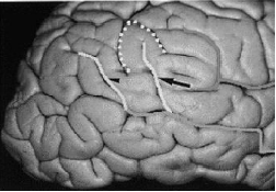
Radiological and Cadaveric Investigation of the Primary Human Brain Sulci: Morphometric Analysis and Clinical Consequences
Keywords:
Human Brain Sulci, Morphometric Analysis, Radiological StudyAbstract
Understanding the connections between different parts of the brain and how they work is a significant focus of current human neuroscience studies. Thanks to advancements in anatomical and functional 3D neuroimaging, what seemed like an insurmountable task a decade ago is now within reach. But before we can create a whole map of the human brain, there are a lot of important questions that need answering. To start, there is a lot of variation in brain anatomy and function from one person to the next, which is why spatial registration and normalisation of brain scans from different people is a hot topic. The second point is that the human brain is spatially and temporally organised at multiple levels, from individual synapses to extensive distributed networks. Neuroimagers face a formidable technical difficulty in integrating these different levels, and solving this problem is a challenging theoretical one in neuroscience. Curiously, similar problem has arisen in functional imaging population studies, where averaging functional images of diverse people became necessary because to the low signal-to-noise ratio of positron emission tomography (PET) images. links between the structure and function of the human brain, both at the macroscopic and microscopic levels, to highlight the significance of the connection between the individual's anatomy and function. The requirement for a shared neuroanatomical reference frame is becoming more pressing as database projects incorporate data from cytoarchitectony, electrical stimulation, electrical recordings, and functional imaging methods such as functional magnetic resonance imaging (fMRI), event-related potentials, magnetoencephalography, electroencephalography, and functional magnetic resonance imaging (PET).
Downloads
References
Amunts K, Jäncke L, Mohlberg H, Steinmetz H, Zilles K. Interhemispheric asymmetry of the human motor cortex related to handedness and gender. Neuropsychologia. 2000;38(3):304–312.
Amunts K, Schlaug G, Schleicher A, Steinmetz H, Dabringhaus A, Roland PE, Zilles K. Asymmetry in the human motor cortex and handedness. Neuroimage. 1996;4:216–222.
Amunts K, Weiss PH, Mohlberg H, Pieperhoff P, Eickhoff SB, Gurd JM, Marshall JC, Shah NJ, Fink GR, Zilles K. Analysis of neural mechanisms underlying verbal fluency in cytoarchitectonically defined stereotaxic space— The roles of Brodmann areas 44 and 45. Neuroimage. 2004;22(1):42–56.
Anderson B, Southern BD, Powers RE. Anatomic asymmetries of the posterior superior temporal lobes: A postmortem study. Neuropsychiatry Neuropsychol Behavioral Neurology. 1999;12(4):247–254.
Ashburner J, Friston KJ. Voxel-based morphometry—The methods. Neuroimage. 2000;11:805–821.
Berger MS, Cohen WA, Ojemann GA. Correlation of motor cortex brain mapping data with magnetic resonance imaging. Journal of Neurosurgery. 1990;72:383–387.
Blanton RE, Levitt JG, Thompson PM, Narr KL, Capetillo-Cunliffe L, Nobel A, Singerman JD, McCracken JT, Toga AW. Mapping cortical asymmetry and complexity patterns in normal children. Psychiatry Research. 2001;107(1):29–43.
Broca P. Anatomie comparée des circonvolutions cérébrales: Le grand lobe limbique et la scissure limbique dans la série des mammifères. Revue d’Anthropologie serie 2. 1878;1:384–398.
Brodmann K. Vergleichende Lokalisationslehre der Grosshirnrinde in Ihren Prinzipien Dargestellt auf Grund des Zellenbaues. Leipzig: 1909.
Brodmann K. Localisation in the Cerebral Cortex. London: Smith-Gordon; 1994. 11. Carpenter MB, Sutin J. Human Neuroanatomy. Baltimore: Williams & Wilkins; 1983.
Chi JG, Dooling EC, Gilles FH. Gyral development of the human brain. Annals of Neurology. 1977;1:86–93.
Chi JG, Dooling EC, Gilles FH. Left-right asymmetries of the temporal speech areas of the human fetus. Archives of Neurology. 1977;34:346–348.
Clarke E, Dewhurst K. An Illustrated History of Brain Function. Oxford: Sandford Publications; 1972.
Clarke S,Miklossy J. Occipital cortex in man: Organization of callosal connections, related myelo- and cytoarchitecture, and putative boundaries of functional visual areas. Journal of Comparative Neurology. 1990;298:188–214.
Standring S, editor. Gray’s anatomy e-book: the anatomical basis of clinical practice. Chapter 25 cerebral hemisphere. Elsevier Health Sciences; 2021. p. 373.
Gonul Y, Songur A, Uzun I, Uygur R, Alkoc OA, Caglar V, Kucuker H. Morphometry, asymmetry and variations of cerebral sulci on superolateral surface of cerebrum in autopsy cases. Surg Radiol Anat 2014; 36(7): 651e661.
Lohmann G, Von Cramon DY, Colchester AC. Deep sulcal landmarks provide an organizing framework for human cortical folding. Cerebr Cortex 2008; 18(6): 1415e1420.
Ribas GC, Ribas EC, Rodrigues CJ. The anterior sylvian point and the suprasylvian operculum. Neurosurg Focus 2005; 18(6): 1e6. 5. Yasargil MG, Krisht AF, Tu¨re U, Al-Mefty O, Yasargil DC. Microsurgery of insular gliomas: part Idsurgical anatomy of the sylvian cistern. Contemp Neurosurg 2017; 39(11): 1e8.
Flores LP. Occipital lobe morphological anatomy: anatomical and surgical aspects. Arq Neuropsiquiatr 2002; 60: 566e571. 7. Central nervous system. In: Mc Minn RMH, editor. Last’s anatomy regional and applied. 9th ed.151. London: Churchill Livingstone; 1994. pp. 579e591.
Crossman AR. Cerebral hemishphere. In: Standring S, editor. Gray’s anatomy the anatomical basis of clinical practice. 39th ed.287. Elsevier; 2005. pp. 388e389. 403.
Mandal L, Mandal SK, Dutta S, Ghosh S, Singh R, Chakraborty SS. Variation of the major sulci of the occipital lobee a morphological study. Al Ameen J Med Sci 2014; 7(2): 141e145.
Juch H, Zimine I, Seghier ML, Lazeyras F, Fasel JH. Anatomical variability of the lateral frontal lobe surface: implication for intersubject variability in language neuroimaging. Neuroimage 2005; 24(2): 504e514.
Tomaiuolo F, MacDonald JD, Caramanos Z, Posner G, Chiavaras M, Evans AC, Petrides M. Morphology, morphometry and probability mapping of the pars opercularis of the inferior frontal gyrus: an in vivo MRI analysis. Eur J Neurosci 1999; 11(9): 3033e3046
Crivello F, Tzourio N, Poline JB, Woods RP, Mazziotta JC, Mazoyer B. Intersubject variability in functional anatomy of silent verb generation: Assessment by a new activation detection algorithm based on amplitude and size information. Neuroimage. 1995;2:253–263.
Dammers J, Mohlberg H, Boers F, Tass P, Amunts K, Mathiak K. A new toolbox for combining magnetoencephalographic source analysis and cytoarchitectonic probabilistic data for anatomical classification of dynamic brain activity. Neuroimage. 2007;34:1577–1587.
Desikan RSH, Ségonne F, Fischl B, Quinn B, Dickerson BC, Blacker D, Buckner RL, Dale AM, Maguire RP, Hyman BT, Albert MS, Killiany RJ. An automated labelling system for subdividing the human cerebral cortex on MRI scans into gyral based regions of interest. Neuroimage. 2006; 31:968–980.
Draganski B, Gaser C, Busch V, Schuierer G, Bogdahn U, May A. Neuroplasticity: Changes in grey matter induced by training. Nature. 2004;427(6972):311–312. 538
Ebeling U, Steinmetz H, Huang Y, Kahn T. Topography and identification of the inferior precentral sulcus in MR imaging. American Journal of Neuroradiology. 1989;10:937–942.
Eickhoff SB, Stephan KE, Mohlberg H, Grefkes C, Fink GR, Amunts K, Zilles K. A new SPM toolbox for combining probabilistic cytoarchitectonic maps and functional imaging data. Neuroimage. 2005;25:1325–1335.
Eickhoff SB, Weiss PH, Amunts K, Fink GR, Zilles K. Identifying human parieto-insular vestibular cortex using fMRI cytoarchitectonic mapping. Human Brain Mapping. 2006;27:611–621.
Fox PT, Burton H, Raichle ME. Mapping human somatosensory cortex with positron emission tomography. Journal of Neurosurgery. 1987;67:34–43.
Friston KJ, Holmes AP, Worsley KJ, Poline JB, Frith CD, Frackowiak RSJ. Statistical parametric maps in functional imaging: A general approach. Human Brain Mapping. 1995;2:189–210.
Friston KJ, Passingham RE, Nutt JG, Heather JD, Sawle GV, Frackowiak RSJ. Localisation in PET images: Direct fitting of the intercommissural (AC-PC) line. Journal of Cerebral Blood Flow & Metabolism. 1989;9:690–695.
Galaburda AM. La région de Broca: Observations anatomiques faites un siècle après la mort de son découvreur. Revue Neurologique. 1980;136(10):609–616.
Galaburda AM, LeMay M, Kemper TL, Geschwind N. Right-left asymmetries in the brain. Science. 1978; 199: 852–856.
Galaburda AM, Sanides F. Cytoarchitectonic organization of the human auditory cortex. Journal of Comparative Neurology. 1980;190:597–610.
Galuske RAW, Schlote W, Bratzke H, Singer W. Interhemispheric asymmetries of the modular structure in human temporal cortex. Science. 2000;289(5486): 1946–1949.
Gannon PJ, Holloway RL, Broadfield DC, Braun AR. Asymmetry of chimpanzee planum temporale: Humanlike pattern of Wernicke’s brain language area homolog. Science. 1998;279(5348):220–222.
Gaser C, Schlaug G. Brain structures differ between musicians and non-musicians. Journal of Neurosciences. 2003;23(27):9240–9245.
Geschwind N, Levitsky W. Human brain left-right asymmetries in temporal speech region. Science. 1968;161: 186–187.
Good C, Johnsrude IS, Ashburner J, Henson RNA, Friston KJ, Frackowiak RSJ. A voxel-based-morphometry study of ageing in 465 normal adult human brains. Neuroimage. 2001;14:21–36.
Grachev ID, Berdichevsky D, Rauch SL, Heckers S, Kennedy DN, Caviness VS, Alpert NM. A method for assessing the accuracy of intersubject registration of the human brain using anatomical landmarks. Neuroimage. 1999;9:250–268.
Hagmann P, Cammoun L, Martuzzi R, Maeder P, Clarke S, Thiran JP, Meuli R. Hand preference and sex shape the architecture of language networks. Human Brain Mapping. 2006;27(10):828–835. 37.
Lemaître H, Crivello F, Grassiot B, Alpérovitch A, Tzourio C, Mazoyer BM. Age- and sex-related effects on the neuroanatomy of healthy elderly. Neuroimage. 2005; 26:900–911.
Mangin JF, Rivière D, Cachia A, Duchesnay E, Cointepas Y, Papadopoulos-Orfanos D, Scifo P, Ochiai T, Brunelle F, Régis J. A framework to study the cortical foldings patterns. Neuroimage. 2004;23:S129–S138.
Mazziotta JC, Toga AW, Evans AC, Fox PT, Lancaster JL, Zilles K, Woods RP, Paus T, Simpson G, Pike B, Holmes CJ, Collins L, Thompsom PJ, MacDonald D, Iacoboni M, Schormann T, Amunts K, PalomeroGallagher N, Geyer S, Parsons LM, Narr KL, Kabani N, LeGoualher G, Boomsma DI, Cannon TD, Kawashima R, 31 Biological Underpinnings of Anatomic Consistency and Variability in the Human Brain 539 Mazoyer BM. A probabilistic atlas and reference system for the human brain: International Consortium for Brain Mapping (ICBM). Philosophical Transactions Royal Society London B. 2001;356:1293–1322.
McRae DL, Branch CL, Milner B. The occipital horns and cerebral dominance. Neurology. 1968;18:95–98.
Nieuwenhuys R, Voogd J, van Huijzen C. The Human Central Nervous System. A Synopsis and Atlas. 3rd ed. Berlin: Springer-Verlag, 1988.
Ochiai T, Grimault S, Scavarda D, Roch G, Hori T, Riviere D, Mangin JF, Regis J. Sulcal pattern and morphology of the superior temporal sulcus. Neuroimage. 2004; 22(2):706–719.
Ono M, Kubik S, Abernathey CD. Atlas of the Cerebral Sulci. New York: Thieme Medical Publishers, Inc.; 1990.
Parker GJ, Luzzi S, Alexander DC, Wheeler-Kingshott CA, Ciccarelli O, Lambon Ralph MA. Lateralization of ventral and dorsal auditory-language pathways in the human brain. Neuroimage. 2005;24(3):656–666.
Paus T, Tomaiuolo F, Otaky N, MacDonald D, Petrides M, Atlas J, Morris R, Evans AC. Human cingulate and paracingulate sulci: Pattern, variability, asymmetry, and probabilistic map. Cerebral Cortex. 1996;6:207–214.
Penhune VB, Zatorre RJ, MacDonald D, Evans AC. Interhemispheric anatomical differences in human primary auditory cortex: Probabilistic mapping and volume measurement from magnetic resonance scans. Cerebral Cortex. 1996;6(5):661–672.
Rademacher J, Caviness VS, Steinmetz H, Galaburda AM. Topographical variation of the human primary cortices: Implications for neuroimaging, brain mapping, and neurobiology. Cerebral Cortex. 1993;3:313–329.
Rajkowska G, Goldman-Rakic PS. Cytoarchitectonic definition of prefrontal areas in the normal human cortex: I. Remapping of areas 9 and 46 using quantitative criteria. Cerebral Cortex. 1995;5:307–322. 58. Roland PE, Zilles K. Brain atlases—A new research tool. Trends in Neurosciences. 1994;17(11):458–467.
Roland PE, Zilles K. Structural divisions and functional fields in the human cerebral cortex. Brain Research Reviews. 1998;26(2–3):87–105.
Rumeau C, Tzourio N, Murayama N, Peretti-Vitton P, Levrier O, Joliot M, Mazoyer B, Salamon G. Location of hand function in the sensorimotor cortex: Magnetic resonance and functional correlation. American Journal of Neuroradiology. 1994;15:425–433.
Salamon G, Gelbert F, Alicherif A, Poncet M, Khalil R, Sobel D,Von Einseidel O,Morel M,Corbaz JM. Le repérage in vivo des aires du langage. Rev Neurol. 1987;143: 580–587.
Sastre-Janer F, Regis J, Belin P, Mangin J-F, Dormont D, Masure MC, Remy P, Frouin V, Samson Y. Three dimensional reconstruction of the human central sulcus reveals a morphological correlates of the hand area.Cerebral Cortex. 1999;8:641–647.
Schleicher A, Amunts K, Geyer S, Morosan P, Zilles K. Observer-independent method for microstructural parcellation of cerebral cortex: A quantitative approach to cytoarchitectonics. Neuroimage. 1999;9:165–177. 64. Schormann T, Zilles K. Three-dimensional linear and nonlinear transformation: An integration of light microscopical and MRI data. Human Brain Mapping. 1998;6: 339–347.
Sluming V, Barrick T, Howard M, Cezayirli E, Mayes A, Roberts N. Voxel-based morphometry reveals increased gray matter density in Broca’s area in male symphony orchestra musicians. Neuroimage. 2002;17(3): 1613–1622.
Sobel DF, Gallen CC, Schwartz BJ, Waltz TA, Copeland B,Yamada S,Hirschkoff EC, Bloom FE. Locating the central sulcus: Comparison of MR anatomic and magnetoencephalographic functional methods. AJNR. 1993; 14:915–925.
Sowell ER, Thompson PM, Holmes CJ, Jernigan TL, Toga AW. In vivo evidence for post-adolescent brain maturation in frontal and striatal regions. Nature Neuroscience. 1999;2(10):859–861.
Sowell ER, Thompson PM, Rex D, Kornsand D, Tessner KD, Jernigan TL, Toga AW. Mapping sulcal pattern asymmetry and local cortical surface gray matter distribution in vivo: Maturation in perisylvian cortices. Cerebral Cortex. 2002;12(1):17–26.
Talairach J, Szikla G, Tournoux P. Altas d’Anatomie Stéréotaxique du Téléencéphale. Paris: Masson, 1967.
Talairach J, Szikla G, Tournoux P, Prossalentis A, BordasFerrer M, Covello L, Iacob M, Mempel E. Atlas d’Anatomie Stéréotaxique du Télencéphale. Paris:1967.
Talairach J, Tournoux P. Co-planar Stereotaxic Atlas of the Human Brain. 3-Dimensional Proportional System: An Approach to Cerebral Imaging. Stuttgart-New York: 1988.
Talairach J, Tournoux P. Co-planar Stereotaxic Atlas of the Human Brain. 3-Dimensional Proportional System: An Approach to Cerebral Imaging. Stuttgart: Georg Thieme Verlag, 1988. 540 Handbook of Medical Image Processing and Analysis
Thompson PM, Schwartz C, Lin RT, Khan AA, Toga AW. Three-dimensional statistical analysis of sulcal variability in the human brain. Journal of Neurosciences. 1996;16:4261–4274.
Watson JDG, Myers R, Frackowiak RSJ, Hajnal JV, Woods RP, Mazziotta JC, Shipp S, Zeki S. Area V5 of the human brain: Evidence from a combined study using positron emission tomography and magnetic resonance imaging. Cerebral Cortex. 1993;3:79–94.
Weinberger DR, Luchins DJ, Morihisia J, Wyatt RJ. Asymmetrical volumes of the right and left frontal and occipital regions of the human brain. Annals of Neurology. 1982;11:97–100.
Wohlschläger AM, Specht K, Lie C, Mohlberg H, Wohlschläger A, Bente K, Pietrzyk U, Stöcker T, Zilles K, Amunts K, Fink GR. Linking retinotopic fMRI mapping and anatomical probability maps of human occipital areas V1 and V2. Neuroimage. 2005;26:73–82.

Downloads
Published
How to Cite
Issue
Section
License

This work is licensed under a Creative Commons Attribution 4.0 International License.
Current Clinical and Medical Education


