Strongyloidiasis: A Neglected Disease but Highly Fatal
Keywords:
Strongyloidiasis, disseminated disease, Strongyloid stercoralis, hyper infection, immunosuppressive patientsAbstract
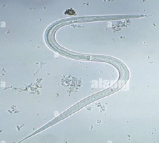
Strongyloidiasis is commonly refer to helminthic infection by a small size parasite belongs to intestinal nematodes named Strongyloid stercoralis which is most common helminthic parasite of humans and most common in tropic and subtropic arias. The disease is world wide distribution and estimated 300 to 600 million individuals around Africa, Asia, South and central America. Bad sanitation, poor hygiene, rural regions and bad socioeconomically status, these are important factor for transmitted the infection. The transmission mainly occurs via attach with fecal contaminated soil. Life cycle of parasite consists of 3 stages they are adult, larvae and egg stage. larvae(L3) serve as infective stage and enter host via penetration of skin. Pathogenicity of parasite occurs due to damage of intestinal mucosa by adult worm also larva migration though body of final host may be cause of many symptoms such as cutaneous, pulmonary, and intestinal symptoms. The microscopic examination of stool, sputum and duodenal content are considered a good method for detect rhabditiform larvae and occasionally filariform larvae. Strongyloidiasis can be treated by both albendazole and ivermectin which are good medications against parasite. long duration of disease or chronic infection is very common and lead to a wide range of clinical manifestations. Ther is two defining features of Strongyloid stercoralis first one the autoinfection which lead to long duration of infection and may lead to hyper infection and finally distribution of parasite through the body and cause disseminated disease usually occurs among immunosuppressive patients. with more than sixty percentage fatality. Prevent and control of strongyloidiasis may be comprised by reduce contamination of soil with stool of humans, sanitary disposal of human feces, wearing shoes and gloves is necessary when contact with infective soil, treatment of all infected cases are effective ways to prevent occurrence of strongyloidiasis.
Downloads
References
Olsen A, van Lieshout L, Marti H, et al. Strongyloidiasis – the most neglected of the neglected tropical diseases? Trans R Soc Trop Med Hyg. 2009;103:967–972.
Ashford RW, Barnish G, Viney ME. Strongyloides fuelleborni kellyi: infection and disease in Papua New Guinea. Parasitol Today. 1992; 8(9):314–318.
Abrescia FF, Falda A, Caramaschi G, et al. Reemergence of strongyloidiasis, Northern Italy. Emerg Infect Dis. 2009;15:1531–1533.
Adams M, Page W, Speare R. Strongyloidiasis: an issue in aboriginal communities. Rural Remote Health. 2003;3(1):152.
Carvalho M, Porto DF. Epidemiological and clinical interaction between HTLV-1 and Strongyloides stercoralis. Parasite Immunol. 2004;26:487–497.
Toledo R, Muñoz-Antoli C, Esteban JG. Strongyloidiasis with emphasis on human infections and its different clinical forms. Adv Parasitol. 2015;88:165–241.
Schär F, Trostdorf U, Giardina F. Strongyloides stercoralis: global distribution and risk factors. PLoS Negl Trop Dis. 2013;7(7):e2288.
Roberts AL, Schneider AE, Young RL, Hinrichs SH, Iwen PC. Strongyloides stercoralis infection in a non-endemic area. Lab Medicine. 2013;44:339–343.
Uparanukraw P, Phongsri S, Morakote N. Fluctuation of larval excretion in Strongyloides stercoralis infection. Am J Med Hyg. 1999;60:967–973.
Scowden EB, Schaffner W, Stone WJ. Overwhelming strongyloidiasis: an unappreciated opportunistic infection. Medicine (Baltimore). 1978;57(6):527–544.
Bur Kukuruzovic R, Robins-Browne RM, Anstey NM, Brewster DR. Enteric pathogens, intestinal permeability and nitric oxide production in acute gastroenteritis. Paediatr Infect Dis J. 2002;21:730–739. ke JA. Strongyloidiasis in childhood. Am J Dis Child. 1978;132:1130–1136.
Fisher D, McCarry F, Currie B. Strongyloidiasis in the Northern Territory. ‘Under-recognised and under-treated?’. Med J Aust. 1993;159:88–90.
Dawson-Hahn EE, Greenberg SL, Domachowske JB, Olson BG. Eosinophilia and the seroprevalence of schistosomiasis and strongyloidiasis in newly arrived pediatric refugees: an examination of Centers for Disease Control and Prevention Screening Guidelines. J Pediatr. 2010;156:1016–1018.
Prociv P, Luke R. Observations on strongyloidiasis in Queensland aboriginal communities. Med J Aust. 1993;158:160–163.
Neumann I, Ritter R, Mounsey A. Strongyloides as a cause of fever of unknown origin. J Am Board Nwokolo C, Imobiosen EAE. Strongyloidiasis of respiratory tract presenting as asthma. Br Med J. 1973;2:153–154.Fam Med. 2012;25(3):390–393.
Woodring JH, Halfhill H 2nd, Reed JC. Pulmonary strongyloidiasis: clinical and imaging features. AJR Am J Roentgenol. 1994;162(3):537–542.
Keiser PB, Nutman TB. Strongyloides stercoralis in the immunocompromised population. Clin Microbiol Rev. 2004;17(1):208–217.Evering T, Weiss LM. The immunology of parasite infections in immunocompro-mised hosts. Parasite Immunol 2006;28:549–65
Chu E, Whitlock WL, Dietrich RA. Pulmonary hyperinfection syndrome with Strongyloides stercoralis. Chest. 1990;97:1475–1477.
Mota-Ferreira DM, Gonçalves-Pires MR, Júnior AF, Sopelete MC, Abdallah VO,Costa-Cruz JM. Specific IgA and IgG antibodies in paired serum and breast milksamples in human strongyloidiasis. Acta Trop 2009;109(2):103–7
Mukerjee CM, Carrick J, Walker JC, Woods RL. Pulmonary strongyloidiasis presenting as chronic bronchitis leading to interlobular septal fibrosis and cured by treatment. Respirology. 2003;8(4):536–540.
Downloads
Published
How to Cite
Issue
Section
License

This work is licensed under a Creative Commons Attribution 4.0 International License.
Current Clinical and Medical Education